Pages

Wednesday, August 20, 2014

'Trojan horse' gold nanoparticles treatment could beat brain tumors


A cancer cell containing the nanoparticles. The nanoparticles are
colored green, and have entered the nucleus, which is the area in blue.
Credit: M. Welland
 
A "Trojan horse" treatment for an aggressive form of brain cancer,
which involves using tiny nanoparticles of gold to kill tumour cells,
has been successfully tested by scientists.

The ground-breaking technique could eventually be used to treat glioblastoma
multiforme, which is the most common and aggressive brain tumour in
adults, and notoriously difficult to treat. Many sufferers die within a
few months of diagnosis, and just six in every 100 patients with the
condition are alive after five years.

The research involved engineering nanostructures containing both gold
and cisplatin, a conventional chemotherapy drug. These were released
into tumour cells that had been taken from glioblastoma patients and
grown in the lab.

Once inside, these "nanospheres" were exposed to radiotherapy. This
caused the gold to release electrons which damaged the cancer cell's DNA
and its overall structure, thereby enhancing the impact of the
chemotherapy drug.

The process was so effective that 20 days later, the cell culture
showed no evidence of any revival, suggesting that the tumour cells had
been destroyed.

While further work needs to be done before the same technology can be
used to treat people with glioblastoma, the results offer a highly
promising foundation for future therapies. Importantly, the research was
carried out on cell lines derived directly from glioblastoma patients,
enabling the team to test the approach on evolving, drug-resistant
tumours.

The study was led by Mark Welland, Professor of Nanotechnology and a
Fellow of St John's College, University of Cambridge, and Dr Colin
Watts, a clinician scientist and honorary consultant neurosurgeon at the
Department of Clinical Neurosciences. Their work is reported in the
Royal Society of Chemistry journal, Nanoscale.

"The combined therapy that we have devised appears to be incredibly
effective in the live cell culture," Professor Welland said. "This is
not a cure, but it does demonstrate what nanotechnology can achieve in
fighting these aggressive cancers. By combining this strategy with
cancer cell-targeting materials, we should be able to develop a therapy
for glioblastoma and other challenging cancers in the future."


  diagram showing the composition of the nanosphere. Credit: M. Welland
To date, glioblastoma multiforme (GBM) has proven very resistant to treatments. One reason for this is that the tumour cells invade surrounding, healthy brain tissue, which makes the surgical removal of the tumour virtually impossible.

Used on their own, chemotherapy drugs
can cause a dip in the rate at which the tumour spreads. In many cases,
however, this is temporary, as the cell population then recovers.

"We need to be able to hit the cancer cells directly with more than
one treatment at the same time" Dr Watts said. "This is important
because some cancer cells are more resistant to one type of treatment
than another. Nanotechnology provides the opportunity to give the cancer
cells this 'double whammy' and open up new treatment options in the
future."

In an effort to beat tumours more comprehensively, scientists have
been researching ways in which gold nanoparticles might be used in
treatments for some time. Gold is a benign material which in itself
poses no threat to the patient, and the size and shape of the particles
can be controlled very accurately.

When exposed to radiotherapy, the particles emit a type of low energy
electron, known as Auger electrons, capable of damaging the diseased
cell's DNA and other intracellular molecules. This low energy emission
means that they only have an impact at short range, so they do not cause
any serious damage to healthy cells that are nearby.

In the new study, the researchers first wrapped gold nanoparticles
inside a positively charged polymer, polyethylenimine. This interacted
with proteins on the cell surface called proteoglycans which led to the
nanoparticles being ingested by the cell.


Once there, it was possible to excite it using standard radiotherapy,
which many GBM patients undergo as a matter of course. This released
the electrons to attack the cell DNA.

While gold nanospheres, without any accompanying drug, were found to
cause significant cell damage, treatment-resistant cell populations did
eventually recover several days after the radiotherapy. As a result, the
researchers then engineered a second nanostructure which was suffused
with cisplatin.

The chemotherapeutic effect of cisplatin combined with the radiosensitizing effect of gold nanoparticles
resulted in enhanced synergy enabling a more effective cellular damage.
Subsequent tests revealed that the treatment had reduced the visible
cell population by a factor of 100 thousand, compared with an untreated
cell culture, within the space of just 20 days. No population renewal
was detected.

The researchers believe that similar models could eventually be used
to treat other types of challenging cancers. First, however, the method
itself needs to be turned into an applicable treatment for GBM patients.
This process, which will be the focus of much of the group's
forthcoming research, will necessarily involve extensive trials. Further
work needs to be done, too, in determining how best to deliver the
treatment and in other areas, such as modifying the size and surface
chemistry of the nanomedicine so that the body can accommodate it
safely.

Sonali Setua, a PhD student who worked on the project, said: "It was
hugely satisfying to chase such a challenging goal and to be able to
target and destroy these aggressive cancer cells.
This finding has enormous potential to be tested in a clinical trial in
the near future and developed into a novel treatment to overcome
therapeutic resistance of glioblastoma."

Welland added that the significance of the group's results to date
was partly due to the direct collaboration between nanoscientists and
clinicians. "It made a huge difference, as by working with surgeons we
were able to ensure that the nanoscience was clinically relevant," he
said. "That optimises our chances of taking this beyond the lab stage,
and actually having a clinical impact."

Tuesday, August 19, 2014

Steroids vs Dietary Therapy to Treat Eosinophilic Esophagitis

http://www.mclno.org/webresources/kbase/cellatlas/cell%20images/Eosinophil.jpg


Abstract and Introduction

Abstract

Purpose of Review. Eosinophilic esophagitis (EoE) is a condition characterized by dense mucosal eosinophilia
in conjunction with symptoms of esophageal dysfunction. Since both the
incidence and prevalence of EoE are on the rise in both children and
adults, understanding the various treatment options available is
imperative in choosing the proper treatment for each patient. This
article will highlight the major strides in both medical and dietary
treatment of EoE in the past year.


Recent Findings. Whereas prior
studies have shown that medical therapy with topical corticosteroids is
effective in treating EoE, this more recent literature highlights some
of the limitations of this approach, raising awareness that development
of better drug delivery models is greatly needed. The review also
describes the recent advances in the field of dietary therapy for this
disease, particularly in adults, and further supports the notion that
the pathophysiology of this disease in children and adults is similar,
with food antigens driving this disease.


Summary. Both medical and dietary
therapy are effective for treating adults and children with EoE.
Choosing the optimal treatment approach should be individualized based
both on patient goals and on available local resources. Future
prospective clinical trials comparing these two treatment modalities are
needed to help understand comparable effectiveness as well as to help
understand potential predictors of response to treatment and identify
optimal therapeutic endpoints.


Introduction

Recent consensus guidelines define
eosinophilic esophagitis (EoE) as a chronic, immune/antigen-mediated
esophageal disease characterized clinically by symptoms related to
esophageal dysfunction and histologically by eosinophil-predominant
inflammation.[1] The most common symptoms of adults with this condition include dysphagia and food impaction.[1,2]
Involvement of allergic mechanisms in the pathogenesis of EoE has been
supported by prior studies demonstrating esophageal tissue expression of
allergic mediators such as IgE, eotaxin-3, IL-13, IL-5 and esophageal
involvement by immune cells, including mast cells, dendritic cells and
eosinophils.[3] Furthermore, esophageal eosinophilia is induced in a murine model following allergen exposure.[4]
Given the immunological reactivity of the disease, treatment with
anti-inflammatory medications such as oral corticosteroids and swallowed
topical corticosteroids has been shown to be efficacious in both
children and adults.[5–8]


The concept of food allergens as the main antigenic trigger in EoE
was introduced in a landmark study by Kelly and Sampson in pediatric
patients with symptoms of gastroesophageal reflux disease (GERD) and
histologic features of esophageal eosinophilia, both of which were
unresponsive to acid suppression or fundoplication. After treatment with
an elemental or amino acid based formula, both symptoms and histologic
eosinophilia resolved.[9,10]
Since this landmark study, numerous series have replicated this
association of food allergens as a trigger in EoE in both the adult and
the pediatric population.[2,5,11–13]
Common food triggers found to cause EoE in both children and adults
include milk, wheat, soy, egg, nuts/peanuts, and fish/shellfish.[11,12]


The feasible goals of treatment in EoE are still evolving, but
typically include resolution of clinical symptoms, and the achievement
and maintenance of histologic remission. Other important goals include
prevention of complications of the disease (including fibrostenotic
changes such as strictures), avoidance of food bolus impaction and
avoidance of esophageal perforation, which can occur either
spontaneously (from retching during a food impaction) or iatrogenically
(from stricture dilation). Other important therapeutic endpoints include
improvement in the patient's quality of life, improvement of
nutritional deficits in those treated with dietary restrictions, and
prevention of harmful side effects of medications used to treat the
disease. This study highlights the recent strides in both dietary and
medical therapy over the past year.


Comparison of Different Formulations of Topical Corticosteroids

As mentioned, few studies have tried to
identify differences in the effectiveness of EoE medications based on
changes in their formulation or methods of drug delivery. Dellon et al.[15] performed a randomized trial comparing nebulized and viscous topical steroid preparations in a cohort of adult patients.
Patients received budesonide 1 mg twice daily, either in an aerosolized
form swallowed from a nebulizer or as an oral viscous slurry, for a
total of 8 weeks. Study endpoints included dysphagia improvement based
on MDQ score, reduction in eosinophil counts, and mucosal medication
contact time, which was measured by nuclear scintigraphy with tagged
radiocontrast. The authors found that histologic improvement was
significantly higher in the oral viscous budesonide group (64%) than in
the swallowed nebulizer solution group (27%), although both groups had
comparable improvement in their dysphagia scores. Nuclear scintigraphy
showed that mucosal contact time was much higher in patients treated
with the oral viscous budesonide than the nebulizer solution. This study
showed that the frequency of histologic improvement may be directly
related to mucosal contact time, and has highlighted the importance of
appropriate drug delivery methods in treatment of this disease. It also
shows that complete histologic resolution was achieved in only 64% of
the oral viscous budesonide group, which is a lower percentage than has
been found in prior studies.


Topical Corticosteroids Versus Acid Suppression

In the past year, several studies have
confirmed the effectiveness of medical therapy for EoE, but also have
highlighted some limitations of the current treatment approach. Moawad et al.[14] recently performed a randomized controlled trial of swallowed fluticasone versus esomeprazole for the treatment of esophageal eosinophilia.
In this study, 42 patients suspected of having EoE (defined by clinical
symptoms of esophageal dysfunction and >15 eos/hpf) underwent
esophageal pH testing with 24 h pH/impedance monitoring. Patients were
then stratified by the presence of GERD and randomized to receive either
fluticasone 440 μg twice daily or esomeprazole 40 mg once daily for 8
weeks. Repeat endoscopy was performed, and the primary outcome was a
histologic response of less than 7 eos/hpf. Secondary outcomes included
clinical change in symptoms using the Mayo Dysphagia Questionnaire (MDQ)
score and interval change in endoscopic features. The investigators
showed that there was no difference in the frequency of resolution of
esophageal eosinophilia between the fluticasone and esomeprazole groups
(19 versus 33%; P = 0.484). They also found that patients with
established GERD (based on pH testing) were much more likely to respond
to proton pump inhibitor (PPI) than fluticasone. Interestingly, they
also found that symptoms improved with PPI treatment, but not with
fluticasone. They concluded that the histologic response with both
treatment options was similar and significantly less than expected
(based on prior studies). This study raises concern about the limited
efficacy of some formulations of swallowed aerosolized topical
corticosteroids, and highlights the importance of using acid suppression
as part of the initial diagnostic and therapeutic strategy for patients
with esophageal symptoms and eosinophilia.


Medical Therapy in Eosinophilic Esophagitis

While prior studies have shown the efficacy
of both systemic and topical corticosteroids in the treatment of EoE,
currently there is no medication specifically approved by the US Food
and Drug Administration (US FDA) for the disease. There have also been
few studies comparing the effectiveness of different types of
pharmacologic therapies. The two most common steroid medications used
for treatment of EoE have been fluticasone and budesonide. Fluticasone
is typically delivered via a metered-dose inhaler whereby the medication
is puffed into the mouth and then swallowed. Starting doses in
published studies have ranged from 440 to 880 μg b.i.d. Budesonide has
been used in the form of oral viscous budesonide, which is a suspension
of the drug in a sucralose binder. Patients are typically counseled on
mixing this medication with multiple packets of sucralose to make a
viscous solution that is swallowed twice daily. A typical starting dose
in adults is 1 mg twice daily.


Placebo-controlled Trials of Swallowed Topical Corticosteroids

Other adult studies also have shown limitations with the use of swallowed topical corticosteroids. For example, Alexander et al.[16]
performed a double-blind, randomized, placebo-controlled trial of
fluticasone in 42 adult patients with EoE. Patients in the treatment arm
swallowed 880 μg of aerosolized fluticasone (four puffs) twice daily
for 6 weeks. Their therapeutic endpoints were symptomatic (based on the
MDQ-30) and histologic response (based on a criteria of >90% decrease
in mean eosinophil count). They reported complete histologic response
in 11/15 (62%) patients receiving 6 weeks of fluticasone compared with
none of the 15 patients receiving placebo (P < 0.001, based
on intention-to-treat analysis). Dysphagia was reduced in only 57% of
patients receiving fluticasone compared with 33% receiving placebo
therapy (P = 0.22 by intention-to-treat analysis), and esophageal candidiasis
was found in 26% of patients treated with fluticasone. These results
highlight the fact that topical corticosteroids result in less
histologic response than suggested by earlier studies, and have higher
rates of important side effects, including candidiasis, than previously
reported. As in the prior study, the decreased histologic response is
likely due to problems in drug delivery with attempting to swallow an
aerosolized medication, as well as insufficient mucosal contact time.


These recent pharmacologic studies in EoE demonstrate that medical
therapy can result in histologic remission in some patients, but the
limitations shown by the above studies highlight the important unmet
need to identify better medications for the treatment of EoE.


Dietary Therapy in Eosinophilic Esophagitis

Dietary therapy has long been accepted as
first-line therapy in children with EoE, and recently has been shown to
be effective in adults as well. Three approaches to dietary elimination
in EoE patients have evolved. The first is total elimination of all food
allergens by placing patients on an elemental or amino acid-based
formula as their primary nutrition. Patients are usually placed on this
diet for at least 6 weeks. Another approach to dietary therapy has been
allergy-directed diets using the information gained from allergy testing
(e.g. skin prick) to help guide the foods that are to be restricted.
While this approach has been shown to be helpful in some pediatric
cohorts,[11]
it had limited utility in adult populations due to the lack of
correlation between foods identified by allergy testing and food
triggers in individual patients.[1,2,13,17]
The last approach of empiric dietary elimination (e.g. six-food
elimination diet) has been shown to be equally effective in children and
adults.[2,12]


Goals of Diet Therapy

Knowing that dietary therapy is effective in
adults provides the rationale for offering this treatment approach as an
alternative to swallowed topical corticosteroids. It is important to
emphasize that the goals of the dietary therapy are not to stay on a
restrictive diet indefinitely, but, rather, to undergo a process of food
trigger identification to help tailor the diet for long-term
maintenance therapy. Dietary elimination with serial food reintroduction
enables the identification of the actual food triggers of the disease.
This treatment plan should be individualized based on individual
patients and their goals.


Effectiveness of Empiric Elimination Diets in Adults

While dietary therapy has been well
established as an effective first-line therapy in pediatrics for EoE,
this approach has not been extensively studied in adults. Recently,
empiric dietary elimination has been shown to have comparable
effectiveness in adults. Gonsalves et al. prospectively studied
the efficacy of the six-food elimination diet (SFED) in 50 adults (25
M/25 F) with EoE. Seventy percent of patients had histologic response of
less than 10 eos/hpf, 94% had symptomatic improvement and 74% had
endoscopic improvement after completing the diet for 6 weeks. Serial
food reintroduction was undertaken in patients who responded to the
diet. When food triggers were identified, symptoms typically recurred
within 5 days and esophageal eosinophil counts returned to pretreatment
values (P < 0.0001) on follow-up endoscopy. Common food
allergens identified during this process were wheat (60%), milk (50%),
soy (10%), nuts (10%) and egg (5%). The majority of patients had only
one food trigger. Skin prick testing for food allergens was undertaken
prior to the elimination diet, but was predictive of food triggers in
only 13% of cases.


Subsequent to the publication of this study, Lucendo et al.[13]
from Spain demonstrated similar results in 67 adults with EoE after
empiric elimination of wheat, rice, corn, legumes, peanuts, soy, egg,
milk, fish and shellfish for a similar duration. This approach resulted
in histologic improvement of less than 15 eos/hpf in 73% of the
patients, but required additional foods to be removed.[11]
Food reintroduction in this study identified the common triggers as
milk (61%), wheat (28%), eggs (26%) and legumes (23%). Unlike the prior
study, the majority of patients in this study were found to have
multiple food antigens as their triggers. A single offending food
antigen was identified in only 36%, two food triggers were found in 31%,
and three or more triggers were found in 33% of patients. Results of
allergy testing in this cohort were also not predictive of their food
triggers. The investigators also found that continued elimination of the
food triggers was effective in maintaining remission.


Noting that milk was one of the most common food triggers in their pediatric cohort, Kagalwalla et al.[18]
investigated the utility of a single food elimination diet in their EoE
patients. In this retrospective study conducted between 2006 and 2011,
17 patients who had empirically eliminated cow's milk protein from the
diet and had a follow-up endoscopy were included. Sixty-five per cent of
these patients achieved remission as defined by less than 15 eos/hpf
after therapy.[18]
Although this study has encouraging results, further prospective
studies will be needed in both pediatrics and adults to assess the
generalizability of the approach of single food elimination.


Effectiveness of Allergy-directed Diets in Adults

One of the first studies to attempt a form of
allergy-directed diet in adults was pursued in a small number of adult
patients in Switzerland by Simon et al.[19]
In this study, based on results of IgE testing to certain foods, few
foods were eliminated from the diet. Despite elimination of these foods,
patients did not respond symptomatically. The one patient who completed
endoscopic evaluation after dietary therapy also did not show a
histologic response. A more recent study undertaken by Molina-Infante et al.[20]
studied the outcome of an allergy-directed diet using a multimodal
approach including skin prick testing, prick-prick testing and atopy
patch testing to identify allergens in their adult EoE cohort of 22
patients. Disappointingly, they found only a 26% improvement with this
approach. Likewise, allergy testing was not found to be predictive of
food triggers in either the Gonsalves or the Lucendo study. Taken
together, these studies suggest that current allergic testing methods
are not reliable for identifying food triggers in adults with EoE and
should not be used to guide dietary intervention.


Effectiveness of Elemental Diets in Adults

A recent study by Peterson et al.[21]
evaluated the effectiveness of an elemental diet in a small group of
adults. They found that 50% of adults responded histologically to the
diet with eosinophil counts less than 5 eos/hpf, and 72% had less than
10 eos/hpf, with eosinophil levels on average dropping from 54 to 10
after the diet. Interestingly, patients did not demonstrate symptomatic
improvement, but that may be due to limitations in the dysphagia
assessment tool used in this study. The authors also suspected that the
limited efficacy of elemental diet in this study compared with pediatric
studies might have resulted from poor adherence to the diet in their
adult patient population.


Potential Development of Tolerance After Dietary Therapy

Once a food trigger is identified in EoE, the
mainstay of therapy is continued avoidance of that particular food.
There have not been systematic studies evaluating recurrent food
challenges and the possible development of tolerance over time. One of
the first studies to address this issue was by Leung et al.,[22]
who performed a retrospective review of their pediatric EoE patients
who were known to have milk as their food trigger. They identified 15
patients who had subsequently reintroduced baked milk back into their
diets for at least 6 weeks, and 11 of them (73%) had maintained
histologic remission despite the reintroduction of baked milk products.
The study did not mention the precise time when reintroduction of baked
milk products occurred in these patients. Despite this limitation, this
study suggests that, over time, some patients with cow's milk-induced
EoE may be able to tolerate milk reintroduction in the form of baked
milk, which would allow a considerable broadening of the diet.


Another study exploring this concept was performed by Lucendo et al.[23] in adults. This group evaluated the use of a cow's milk-based hydrolyzed formula in patients with cow's milk-induced EoE.[23]
Seventeen adult patients with cow's milk-induced EoE were administered
this formula over a period of 8 weeks, and repeat endoscopy was
performed. At that time, 88% of patients maintained histologic remission
as defined by less than 15 eos/hpf. This study suggests that some
patients with EoE triggered by a cow's milk protein may tolerate
reintroduction of milk in this reduced antigenic state. While these
formulas are not readily available, this study does provide insight into
the pathophysiologic mechanisms of food allergy that trigger EoE, and
suggests that some patients are tolerant to less antigenic preparations
of their food triggers.


Potential Advantages of Medical Therapy in Adults

Medical therapy has been shown to effectively
induce both symptomatic and histologic remission in some patients with
EoE. Advantages of this approach include the ease of administration of
the medications as well as the limited impact of this treatment approach
on lifestyle. This treatment plan allows patients to eat normally, and
does not mandate dietary restrictions. While an endoscopy after
treatment institution often is performed to ensure that the medication
is working, the medical treatment approach does not require multiple
additional endoscopies, as are often performed in dietary therapy.
Medical therapy does, however, require chronic daily use of medications
with potential side effects.


Potential Advantages of Dietary Therapy

Dietary therapy in adults with EoE has a
number of practical advantages. As prior studies have shown,
discontinuation of swallowed topical corticosteroids in the treatment of
EoE can cause symptomatic and histologic recurrence which may
necessitate chronic daily therapy. Avoidance of food allergens
eliminates the need for chronic medication/corticosteroids to help
control the disease. Oral and esophageal candidiasis
may occur in 5–30% of patients treated with steroids. Other rare side
effects of topical corticosteroids include growth failure in children,
cataracts and adrenal suppression. Unfortunately, these medications have
not been US FDA-approved to be swallowed, which raises concern about
their long-term safety. In addition, long-term use of these medications
may result in a considerable cost to patients with this chronic
condition.


The effectiveness of elimination diet in adults supports the
conceptual definition that EoE is an antigen or immune-mediated
esophageal disease. Therefore, dietary therapy has the advantage of
getting to the root cause of the disease (i.e. food allergen avoidance)
rather than symptom and histologic control with topical corticosteroids.
Current consensus guidelines suggest continued avoidance of food
allergens in patients who are managed with diet therapy,[1,2,5,11,12]
and a targeted, individualized approach to nutrition therapy is
essential to success. Recent studies have suggested that food
reintroduction with altered forms of the food trigger may induce
tolerance and allow more food products to be added. While repeated
endoscopies often are recommended during the food reintroduction
process, recent studies have suggested that other noninvasive testing
methods may become available soon.[24]


Medical Versus Dietary Therapy: Which to Choose?

When discussing options for therapy with
patients, it is important to review the pros and cons of each treatment
plan and try to identify goals of treatment. It is important to
underscore that the goals of dietary therapy are not to stay on the
elemental diet or SFED indefinitely, but, rather, to undergo a process
of food trigger identification to help tailor the diet for long-term
maintenance therapy. The goals of medical therapy are to help control
symptoms and histology with minimal disruption to daily routine, but
will likely require maintenance therapy to achieve these goals. The
choice on proceeding with medical or dietary therapy should be
individualized based on patient preference as well as available local
resources.


Conclusion

Medical and dietary therapy both are
effective treatments for patients with EoE. While previous studies have
demonstrated the effectiveness of topical corticosteroids, research over
this past year has highlighted limitations of this approach. These
studies have underscored the unmet need for additional therapeutic
options with better drug delivery systems that are specific for EoE.
They have also highlighted the need to better define therapeutic
endpoints (i.e. symptomatic, histologic, or endoscopic response or a
combination of all three). Research is underway to help answer these
basic questions and to develop an important disease activity index
specifically for EoE. Over the past year, several studies have also
highlighted the effectiveness of dietary therapy in adults with EoE, and
have demonstrated that this treatment approach should be considered as
first-line therapy in adults, as it is in children. Knowing that both
medical and dietary therapy is effective provides options for patients,
and highlights the need to individualize treatment plans based on
patient preferences.


Friday, August 15, 2014

CDC Accidentally Ships Deadly Virus, Hopes No One Will Notice

CDC Accidentally Ships Deadly Virus, Hopes No One Will Notice

That wacky CDC is up to its old, potentially fatal-virus-spreading tricks again. But instead of anthrax or dengue,
this time, the Centers for Disease Control brought a deadly strain of
bird flu into its revolving cast of highly contagious characters. While
rushing to get to a meeting, a CDC scientist accidentally tainted a tamer strain of bird flu with a far more deadly one—and then sent it out to another unsuspecting lab. Whoops.

This
most recent set of hijinks took place at CDC Prevention headquarters in
Atlanta in January, when a lab scientist accidentally mixed the two
samples, sending what should have been a benign (at least to
humans) strain of the virus to another lab. Except, you know, it wasn't.
So when that very same virus concoction was given to some unsuspecting
chickens as part of a USDA study in March, and all those chickens
proceeded to immediately die, the USDA officials knew something wasn't
right.

The CDC lab responsible for the deadly mixed sample then confirmed that, yes, that virus was actually wildly dangerous but told, well, no one. No one until June, that is, when a second lab reported a similar problem, and CDC Director Dr. Tom Frieden was finally notified.

Apparently,
the lab scientist who had originally contaminated the sample completed
what should have been 90 minutes of work (with both the tame and deadly
viruses) in 51 minutes, in an attempt to make the noon meeting. Whether
that meeting actually did begin as scheduled, though, remains
inconclusive.

To the CDC's credit, "the viral mix was at all times contained in specialized laboratories and was never a threat to the public," according to an internal report. But then that's what they said last time, too. And the time before that. Here's to hoping Ebola's not next. [AP]

Tuesday, August 12, 2014

Sigmoidoscopy Does Cut Risk of Dying From Colon Cancer: Study

Experts said the study, conducted in Norway and reported in the Aug. 13 issue of the Journal of the American Medical Association, confirms the value of sigmoidoscopy screening.
But
in the United States, where few doctors even perform sigmoidoscopy, the
results are unlikely to make a difference in everyday practice.
"Sigmoidoscopy
is definitely second-best to colonoscopy," said Dr. James Church, a
colorectal surgeon at the Cleveland Clinic in Ohio.
That's mainly
because sigmoidoscopy looks only at the lower portion -- or "left side"
-- of the colon, said Church, who was not involved in the study.
Colonoscopy gives doctors a view of the entire colon.
Both
procedures not only detect cancer but also help prevent it -- by
allowing doctors to remove potentially precancerous growths called
polyps. But colonoscopy can pick up polyps throughout the colon.
"Colonoscopy is not perfect, by any means," Church said. But, he added, "if you don't want to get colon cancer, colonoscopy is the best option."
The
new trial was conducted in Norway, where there is no routine colon
cancer screening, explained lead researcher Dr. Oyvind Holme, of
Sorlandet Hospital in Kristiansand, Norway.
That allowed the
researchers to offer one-time sigmoidoscopy screening to over 20,000 50-
to 64-year-old adults, then compare them with 78,000 people the same
age who were not offered any kind of colon cancer screening.
Half
of the sigmoidoscopy group also received a stool test to look for hidden
blood -- another option for colon cancer screening.
Holme's team
found that people who underwent screening were 27 percent less likely to
die of colon cancer over the next decade, versus the unscreened group.
According to Holme, the findings bolster evidence that sigmoidoscopy is a "valuable tool" for colon cancer screening.
But,
he said, they don't mean that sigmoidoscopy is the best tool. "We did
not compare flexible sigmoidoscopy to other screening methods," Holme
pointed out.
In the United States, experts advise most people to begin colon cancer screening at age 50.
The
U.S. Preventive Services Task Force recommends three choices: an annual
stool test; sigmoidoscopy every five years, along with stool testing
every three years; or colonoscopy every 10 years.
In practice, though, colonoscopy is by far the most common test in the United States.
Sigmoidoscopy
does have a number of advantages over colonoscopy, Holme said. The
pre-exam bowel cleanse is easier to take, and the procedure itself is
faster, does not require sedation and can be done by a primary care
doctor or trained nurse -- whereas colonoscopies are done by
gastroenterologists.
Sigmoidoscopy is also much cheaper: In the United States, it costs about $150 to $300, versus $1,000 or more for a colonoscopy.
But
some of those advantages are not as good as they sound, according to
Church. Since patients are not sedated for sigmoidoscopy, he said, it's
actually more uncomfortable than colonoscopy, for example.
What
this study shows, Church said, is that sigmoidoscopy is better than no
screening -- for preventing cancers on the left side of the colon.
Among
patients who were screened, there were 113 cancers per 100,000 people
each year of the study. That compared with 141 per 100,000 in the
unscreened group. But the difference was mostly seen in cancers in the
left side of the colon.
"For right-sided colon cancer, sigmoidoscopy is, statistically, the same as doing nothing," Church said.
In
the end, questions over how sigmoidoscopy fits into colon cancer
screening could actually be moot in the near future, according to an
editorial published with the study.
New DNA stool tests, which are
much more precise than standard stool tests, have the potential to
reduce routine colonoscopies, writes Dr. Allan Brett, of the University
of South Carolina School of Medicine in Columbia.
"Fecal DNA tests
could change the way we think about stool testing," Church said.
"They're so much better than what we have now."
Just yesterday,
the U.S. Food and Drug Administration approved a DNA-based stool test
that detects colon cancer with more than 90 percent accuracy. Still,
experts said, the test is not intended to replace colonoscopies -- and
any positive result would have to be followed up with a colonoscopy to
confirm the diagnosis.
The hope is that new and better options
will encourage more people to get screened. A recent government study
found that one-third of Americans eligible for colon cancer screening
aren't doing it.

Monday, August 11, 2014

Report: Ebola outbreak started in 2-year-old in Guinea

(CNN) -- The worst outbreak of Ebola, which has
killed 961 people and triggered an international public health
emergency, may have started with a 2-year-old patient in a village in
Guinea.

About eight months ago, the toddler, whom researchers believe may
have been Patient Zero, suffered fever, black stool and vomiting. Just
four days after showing the painful symptoms, the child died on December
6, 2013, according to a report published in The New England Journal of Medicine.

Scientists don't know exactly how the toddler contracted the virus.
Ebola is spread from animals to humans through infected fluids or
tissue, according to the World Health Organization.

"In Africa, infection has been documented through the handling of
infected chimpanzees, gorillas, fruit bats, monkeys, forest antelope and
porcupines," WHO says, though researchers think fruit bats are what
they call the virus's "natural host."

Photos: Ebola outbreak in West Africa
Ebola coverage: informing vs. overhyping
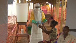
Delayed response cause Ebola to spread?
Man loses 7 relatives to Ebola
Researchers who published the paper this year found a chain of illnesses in the toddler's family.

After the child's death, the mother suffered bleeding symptoms and
died on December 13, according to the report. Then, the toddler's
3-year-old sister died on December 29, with symptoms including fever,
vomiting and black diarrhea. The illness subsequently affected the
toddler's grandmother, who died on January 1, in the family's village of
Meliandou in Guéckédou.

The area in southern Guinea is close to the Sierra Leone and Liberia borders.

The illness spread outside their village after several people attended the grandmother's funeral.

Funerals tend to bring people in close contact with the body. Ebola
spreads from person to person through contact with organs and bodily
fluids such as blood, saliva, urine and other secretions of infected
people. It has no known cure.

READ: 'This is unprecedented'

Two of the funeral attendees appeared to bring back the virus to
their village, and it spread to health care workers and other family
members who took care of infected patients.

"A health care worker from Guéckédou with suspected disease, seems to
have triggered the spread of the virus to Macenta, Nzérékoré, and
Kissidougou in February 2014," stated the report, noting that more
Guinea towns were affected.

Clusters of the disease popped up in early 2014 in these areas, with
the initial patients suffering fever, vomiting and severe diarrhea,
according to the report. Hemorrhaging was less frequent, the report
noted.

In early March, the Ministry of Health in Guinea and Doctors Without Borders in Guinea were notified about the disease clusters.

Health investigators arrived that month and began tracing the disease
by examining hospital documents and conducting interviews with affected
families and villagers.

Ebola has now spread to Liberia, Sierra Leone and Nigeria, prompting global concerns.

The report about the emergence of Ebola in Guinea was authored by
dozens of international doctors and researchers from institutions in
France, Germany, Guinea, WHO and Doctors Without Borders.

 - CNN.com

Sunday, August 10, 2014

PRAGUE CRITERIA - For Endoscopically Suspected Oesophageal Columnar Metaplasia / Barrett’s Oesophagus

In the past, criteria for the diagnosis and grading of the severity of esophageal mucosal consequences of gastroesophageal reflux disease (GERD) have been postulated with insufficient consultation and critical evaluation. The result has been a confusion of inadequately defined criteria, which impairs the quality of communication about endoscopic findings in both routine patient care and research studies. Endoscopists were unable to recognize these changes with acceptable agreement. The result was a lack of consensus on how to assess the presence and extent of CLE in clinical practice. The grading systems which have been proposed previously include:
  1. The classification of CLE into long (≥ 3 cm) and short (<3 cm) segments: this system seems clinically irreverent because it was documented that even SSBE may undergo progression to dysplasia as well as adenocarcinoma.[4-7]
  2. The Z-line appearance (ZAP) classification: this was developed to describe the endoscopic extent of CLE with particular reference to SSBE.[26] The ZAP was found to correlate with the prevalence of IM at the SCJ; however, this system also uses a threshold of 3 cm to distinguish between grades II and III CLE making it insufficiently precise for documenting progression or regression of CLE.
Several years of research and deliberations by the International Working Group For The Classification Of Esophagitis (IWGCO) have led to the development and validation of explicit, consensus-driven criteria for the endoscopic diagnosis and grading of CLE called the Prague C & M Criteria for CLE (Fig. 1).[27] The Prague C & M Criteria is a new grading system for CLE, and it was so named because it was first introduced by the IWGCO at the 11th United European Gastroenterology Week meeting in Prague, Czech Republic in 2004. The purpose of these criteria is to simplify and standardize endoscopic characterization of the extent and length of CLE. The new system emphasizes the use of esophageal landmarks to assess the circumference (C) and maximal (M) extent of the endoscopically visualized CLE segment. The key steps in Prague C & M Criteria are: (i) identify the GEJ as at the tops of the gastric mucosal folds; if hiatus hernia is present, do not confuse with the diaphragmatic hiatal impression for the GEJ; (ii) for circumferential columnar-appearing mucosa above the GEJ, define this extent in centimeters above the GEJ: report as the C-value; and (iii) for any tongue-like areas of columnar-appearing mucosa, measure the maximum extent in centimeters above the GEJ: report as the M-value.[27] The primary validation study for these criteria was published by Sharma et al.[28] in November 2006. The criteria are simple, reliable, reproducible and memorable. It is expected to be a practical clinical tool for characterizing the severity of CLE and for evaluating its progression over time. Now there is a need to build experience in the use of these criteria. As there is no widely used method of grading CLE currently, the Prague C & M Criteria are expected to be used worldwide.[29] The most important weakness of this grading system appears to be in patients with short segment CLE. The kappa measures of interobserver agreement fell markedly when the length of tissue involvement was less than 1 cm.
Prague C & M Criteria for Endoscopically Suspected Esophageal Columnar Metaplasia/Barrett's Esophagus. (Reproduced with permission from the International Working Group for the Classification of Oesophagitis (IWGCO) http://www.iwgco.com )

Wednesday, August 6, 2014

Ebola Virus: How Contagious?

Two Americans stricken with the Ebola
virus amid a record outbreak in West Africa are now being treated in
Atlanta, which has triggered fears of a potential outbreak in the United
States.

Infectious disease experts say they don’t expect that to
happen for several reasons. Ebola is hard to contract, they say, and
good infection-control practices can stop its spread.

What's more, Ebola is much less contagious than many other more common diseases. The virus, much like HIV or hepatitis, is spread through blood or bodily fluids and is not airborne.

Many
factors play into how contagious a disease is thought to be, say Jeff
Duchin, MD, an infectious disease expert at the University of
Washington, Seattle, and Amesh Adalja, MD, an infectious disease expert
at the University of Pittsburgh.

Among those factors:

  • How it’s transmitted (airborne, bodily fluids, other)
  • Infection-control practices in place
  • Extent of contact an infected person has with others
  • Percent of the population that has been vaccinated (if a vaccine exists)
To
gauge how contagious different diseases are, experts take these and
other things into account and estimate the average number of people
likely to catch the illness from a single infected person. They call
this the basic reproductive rate or number. The number is an average, a
scientific guess, experts say, and it is likely to vary from country to
country.

"I would anticipate the reproductive rate for Ebola in the U.S. to be zero," Adalja says.

By comparison, measles, diphtheria, and whooping cough
are all airborne, and they can be transmitted by "just being in
face-to-face contact with an infected patient, without touching them,”
Duchin says. When that person coughs or sneezes, others may become
infected after breathing in the organisms.

Here are the estimated,
overall, basic reproductive rates for Ebola and other infectious
diseases, along with how they're spread.

Measles

Airborne -- reproductive rate 12 to 18

The
measles virus spreads through the air when infected people breathe,
cough, or sneeze. Any person exposed to the virus who is not immune
generally gets the disease. It is less common in countries with good
vaccination coverage.

The virus typically grows in the cells lining the lungs and the back of the throat, leading to fever, runny nose, cough, and an extensive rash.

Measles
was declared eliminated in the U.S. nearly 15 years ago, according to
the CDC. Even so, outbreaks still happen. In 2014, 288 people were
confirmed to have had measles as of May 23. No deaths have occurred this
year in the U.S. from measles.


Pertussis (whooping cough)

Droplets, airborne -- reproductive rate 12 to 17

Whooping
cough is highly contagious. The bacteria that cause the disease attach
to the tiny hair-like extensions (known as cilia) in the respiratory system, leading to the violent coughing that can make it hard to breathe.

It's spread when infected people cough or sneeze and others breathe in the bacteria.

Vaccination
protects people, but outbreaks still happen. In 2012, for instance,
more than 48,000 people with whooping cough were reported to the CDC,
with 20 deaths. Most deaths were in infants younger than 3 months. 
Vaccination should begin at age 2 months, the CDC advises. Adults,
especially those who will be around infants, also need a pertussis
booster.

Diphtheria

Respiratory droplets -- reproductive rate 6 to 7

Diphtheria is an infection caused by bacteria that are spread from person to person, usually by coughing or sneezing.

Diphtheria
is not typically a concern in the U.S., Adalja says, due to routine
vaccination. When it does occur, diphtheria can kill 1 in 10 affected,
according to the CDC.

Polio

Person to person -- reproductive rate 5 to 7

The polio virus spreads from person to person and invades the brain and spinal cord, sometimes leading to paralysis.

The
virus is spread from the stool of an infected person or from droplets
from a sneeze or cough. Toys and other objects contaminated with the
virus can also spread the disease.

Most people infected don't have any symptoms. Others have flu-like symptoms that go away.

Only 1 in 100 people infected develop the weakness or paralysis, according to the CDC.

Of
those people who are paralyzed, up to 10% die when the paralysis
affects the breathing muscles. Vaccination  has wiped out polio from
some, but not all, of the world. Only three countries in the world --
Nigeria, Pakistan, and Afghanistan -- have not stopped the spread of
polio, according to the WHO. Stumbling blocks have included resistance
to vaccinations and the reluctance of some leaders to back vaccination
efforts.

Mumps

Droplets of saliva, mucus; contaminated objects -- reproductive rate 4 to 7

Mumps is a viral illness. It's spread by droplets of mucus or saliva when an infected person coughs, sneezes, or talks.

Vaccination
prevents the disease, but outbreaks have still been seen, including in
the U.S. Most people who get mumps recover fully, the CDC says.

HIV

Sharing needles, sexual contact -- reproductive rate 1 to 4

HIV is the virus that can lead to AIDS. The body can't get rid of the virus, so once infected, a person has HIV for life.

HIV is spread mainly by having sex with or sharing needles or other drug equipment with an infected person.

The
estimated reproductive number, 1 to 4, can vary greatly, Adalja says.
It would typically be much lower if someone infected with HIV is on an
antiretroviral drug, does not inject drugs, and does not take part in
other risky behaviors, he says.

page 3

continued...

AIDS
is still a killer in the U.S. According to the CDC, 15,500 people with
AIDS died in 2010, the latest year for the statistics.

Ebola Virus

Bodily fluids, exposure to contaminated needles and other objects -- reproductive rate 1 to 4

Ebola
was first discovered in 1976. Outbreaks have surfaced from time to time
ever since. Ebola is particularly deadly, though. In the current
outbreak, about 60% of those infected have died, according to the CDC.

Experts believe the virus hosts are animals, probably bats.

While
Ebola and HIV have a similar reproductive number, they are different in
many ways, Duchin says. "HIV and Ebola both are present in the blood,
but the ways they infect cells, where they live in the body, are very
different."

Compared to the airborne organisms spread by casual
contact, "it takes effort to get infected with both of these viruses
[HIV and Ebola]," Adalja says.

© 2014 WebMD, LLC. All rights reserved.

Sunday, August 3, 2014

EASL Recommendations on Treatment of Hepatitis C 2014 .:. Latest news .:. EASL - EUROPEAN ASSOCIATION FOR THE STUDY OF THE LIVER

EASL Recommendations on Treatment of Hepatitis C 2014 .:. Latest news .:. EASL - EUROPEAN ASSOCIATION FOR THE STUDY OF THE LIVER

Guidelines for Colonoscopy Surveillance After Cancer Resection

Guidelines for Colonoscopy Surveillance After Cancer Resection: A Consensus Update by the American Cancer Society and the US Multi-Society Task Force on Colorectal Cancer


Recommendations (Table 1Table
1) on the use of surveillance colonoscopy after resection of
colorectal cancer were produced jointly by the US Multi-Society Task
Force on Colorectal Cancer and the American Cancer Society (ACS). They
constitute the updated recommendations of both organizations. The
rationale for combined guidelines by organizations is discussed in the
accompanying joint recommendations on postpolypectomy surveillance.1Winawer,
S.J., Zauber, A.G., Fletcher, R.H., Stillman, J.S., O’Brien, M.J.,
Levin, B., Smith, R.A., Lieberman, D.A., Burt, R.W., Levin, T.R., Bond,
J.H., Brooks, D., Byers, T., Hyman, N., Kirk, L., Thorson, A., Simmang,
C., Johnson, D., and Rex, D.K. Guidelines for Colonoscopy Surveillance
After Polypectomy (a consensus update by the US Multi-Society Task Force
on Colorectal Cancer and the American Cancer Society) .
Gastroenterology. ;
: 1872–1885

Abstract | Full Text | Full Text PDF | PubMed | Scopus (437)See all References
These guidelines were endorsed by the Colorectal Cancer Advisory
Committee of the ACS and by the governing boards of the American College
of Gastroenterology, the American Gastroenterological Association
Institute, and the American Society for Gastrointestinal Endoscopy.

Table 1Postcancer Resection Surveillance Colonoscopy Recommendations
1.
 Patients with colon and rectal cancer should undergo high-quality
perioperative clearing. In the case of nonobstructing tumors, this can
be done by preoperative colonoscopy. In the case of obstructing colon
cancers, computed tomography colonography with intravenous contrast or
double-contrast barium enema can be used to detect neoplasms in the
proximal colon. In these cases, a colonoscopy to clear the colon of
synchronous disease should be considered 3 to 6 months after the
resection if no unresectable metastases are found during surgery.
Alternatively, colonoscopy can be performed intraoperatively.
2.
 Patients undergoing curative resection for colon or rectal cancer
should undergo a colonoscopy 1 year after the resection (or 1 year
following the performance of the colonoscopy that was performed to clear
the colon of synchronous disease). This colonoscopy at 1 year is in
addition to the perioperative colonoscopy for synchronous tumors.
3.
 If the examination performed at 1 year is normal, then the interval
before the next subsequent examination should be 3 years. If that
colonoscopy is normal, then the interval before the next subsequent
examination should be 5 years.
4. Following the
examination at 1 year, the intervals before subsequent examinations may
be shortened if there is evidence of hereditary nonpolyposis colorectal
cancer or if adenoma findings warrant earlier colonoscopy.1Winawer,
S.J., Zauber, A.G., Fletcher, R.H., Stillman, J.S., O’Brien, M.J.,
Levin, B., Smith, R.A., Lieberman, D.A., Burt, R.W., Levin, T.R., Bond,
J.H., Brooks, D., Byers, T., Hyman, N., Kirk, L., Thorson, A., Simmang,
C., Johnson, D., and Rex, D.K. Guidelines for Colonoscopy Surveillance
After Polypectomy (a consensus update by the US Multi-Society Task Force
on Colorectal Cancer and the American Cancer Society) .
Gastroenterology. ;
: 1872–1885

Abstract | Full Text | Full Text PDF | PubMed | Scopus (437)See all References
5.
 Periodic examination of the rectum for the purpose of identifying local
recurrence, usually performed at 3- to 6-month intervals for the first 2
or 3 years, may be considered after low anterior resection of rectal
cancer. The techniques utilized are typically rigid proctoscopy,
flexible proctoscopy, or rectal endoscopic ultrasound. These
examinations are independent of the colonoscopic examinations described
above for detection of metachronous disease.
Table 2Table 2 summarizes the differences in this guideline from previous guidelines on postcancer resection surveillance colonoscopy.

Table 2Differences Between This Guideline and Previous Guidelines on Postcancer Resection Surveillance Colonoscopy
In
addition to careful perioperative clearing of the colorectum for
synchronous lesions, a colonoscopy is recommended 1 year after surgical
resection because of high yields of detecting early second, apparently
metachronous cancers
Clinicians can consider periodic
examination of the rectum for the purpose of identifying local
recurrence after low anterior resection of rectal cancer.

Candidates for Postcancer Resection Surveillance Colonoscopy

In
general, patients who undergo surgical resection of Stage I, II, or III
colon and rectal cancers or curative-intent resection of Stage IV
cancers are candidates for surveillance colonoscopy. Patients who
undergo curative endoscopic resection of Stage I colon cancers are also
candidates for surveillance colonoscopy. Patients with Stage IV colon or
rectal cancer that is unresectable for cure are generally not
candidates for surveillance colonoscopy because their chance of survival
from their primary cancer is low, and the risks of surveillance
outweigh any potential benefit.


PubMedSee all References
In summary, performance of annual colonoscopy for the purpose of
detecting recurrent disease does not have an established survival
benefit for patients with colorectal cancer. (However, as noted below,
there is a rationale for surveillance of the rectum after resection of
rectal cancer for the detection of local recurrence.) The primary goal
of surveillance colonoscopy after resection of colorectal cancer is the
detection of metachronous neoplasms.


Distinguishing Rectal Cancer Versus Colon Cancer Follow-up

Although
there is no established benefit from endoscopic surveillance for the
purpose of detecting early recurrences of the original cancer, in
clinical practice many clinicians distinguish between rectal and colon
cancer in this regard. The distinction is based on differences in the
rates of local recurrence of rectal vs colon cancer. Specifically, in
the case of colon cancer, recurrence at the anastomosis occurs in only
2%–4% of patients.2Barillari,
P., Ramacciato, G., Manetti, G., Bovino, A., Sammartino, P., and
Stipa, V. Surveillance of colorectal cancer (effectiveness of early
detection of intraluminal recurrences on prognosis and survival of
patients treated for cure) . Dis Colon Rectum. ;
: 388–393

Detection of Metachronous Neoplasms

A second potential
benefit of surveillance colonoscopy is the detection of metachronous
cancers at a surgically curable stage, as well as the prevention of
metachronous cancers via identification and removal of adenomatous
polyps. The incidence of metachronous cancers, the timing at which
metachronous cancers occur, and the stage of these cancers at
presentation or identification by surveillance colonoscopy should
determine the optimal intervals for performance of surveillance
colonoscopy directed toward metachronous disease. The evidence from
published studies of postcancer resection surveillance in colonoscopy
was reviewed to determine what these rates and timing of metachronous
cancers are (Table 3Table 3). Limitations in interpretation of this literature were described earlier.

Table 3Metachronous Cancers in Postcancer Resection Surveillance Colonoscopy Studies
StudyNColonoscopiesMetachronous CRCs (all)Metachronous CRCs (within 24 mo)Dukes’ A or BNumber asymptomaticReoperation for cure
Barillari481
126a96b7
Barrier61c
0


Carlsson12954610NSNSNS
Castells199
0


Chen231
40NS44
Eckardt212
0


Granqvist3906001275d6d10
Green3278
422423NSNS
Juhl13331640444
Khoury389388921NSNSNS
Kjeldsen597
10NSNS88
Kronborg239710434NS4
Makela106
1NSNSNS1
McFarland742370


Obrand444
0


Ohlsson53e
0


Patchett132
2NSNS0NS
Pietra207
1NSNSNSNS
Schoemaker3257338551NS
Skaife611609f51NSNSNS
Stigliano322
50NSNSNS
Togashi341157022917NS22
Weber75197212NS2
Total9029940713757692962
aReport states that “more than one half” arose in first 24 months.
bReports
46 combined local recurrences with metachronous tumors, of which 22
were asymptomatic; number calculated assumes similar proportion for
metachronous cancers.
cSubgroup who underwent perioperative colonoscopy.
dReports
26 combined local recurrences with metachronous tumors, of which 10
were Dukes’ A or B and 14 were asymptomatic; numbers calculated assume
similar proportion for metachronous cancers.
eIntensive surveillance subgroup (control group did not undergo routine colonoscopy).
fTwo patients underwent barium enema for completion of incomplete colonoscopy.
From
2% to 7% of patients with colorectal cancer have 1 or more synchronous
cancers in the colon and rectum at the time of initial diagnosis.3Barrier,
A., Houry, S., and Huguier, M. The appropriate use of colonoscopy in
the curative management of colorectal cancer. Int J Colorectal Dis. ;
: 93–98

Alternatives to Colonoscopy for Surveillance

Colonoscopy
is considered the test of choice for detection of metachronous
neoplasms in the postcancer resection surveillance colonoscopy setting (Table 4Table
4). Double-contrast barium enema was less sensitive than colonoscopy
for large and small polyp detection after resection of adenomas.59National
Polyp Study Work Group, Winawer, S.J., Stewart, E.T., Zauber, A.G.,
Bond, J.H., Ansel, H., Waye, J.D., Hall, D., Hamlin, J.A., Schapiro, M.,
O’Brien, M.J., Sternberg, S.S., and Gottlieb, L.A. A comparison of
colonoscopy and double-contrast barium enema for surveillance after
polypectomy. N Engl J Med. ;
: 1766–1772

CrossRef | PubMed | Scopus (437)See all References

Table 4Additional Recommendations Regarding Postcancer Resection Surveillance Colonoscopy
1. These recommendations assume that colonoscopy is complete to the cecum and that bowel preparation is adequate
2.
 There is clear evidence that the quality of examinations is highly
variable; continuous quality improvement process is critical to the
effective application of colonoscopy in colorectal cancer prevention50Rex,
D., Cummings, O., and Ulbright, T. Coming to terms with pathologists
over colon polyps with cancer or high-grade dysplasia. J Clin
Gastroenterol. ;
:

CrossRef | PubMedSee all References
3. Endoscopists should make clear recommendations to primary care physicians about when the next colonoscopy is indicated
4. Performance of fecal occult blood text is discouraged in patients undergoing colonoscopic surveillance
5.
 Discontinuation of surveillance colonoscopy should be considered in
persons with advanced age or comorbidities (<10 years life
expectancy), according to the clinician’s judgment
6. Surveillance guidelines are intended for asymptomatic people; new symptoms may need diagnostic work-up
7. Chromoendoscopy (dye-spraying) and magnification endoscopy are not established as essential to screening or surveillance
8. Computed tomography colonography (virtual colonoscopy) is not established as a surveillance modality
CT colonography has not been evaluated adequately in the surveillance setting, and results for polyp detection are quite mixed.60Rockey, D., Paulson, E., Favis, W. et al. Multicenter prospective comparison of colon imaging tests. (abstr)Gastroenterology. ;
: A2004

See all References, 61Johnson,
C.D., Harmsen, W.S., Wilson, L.A., McCarty, R.L., Welch, T.J., Ilstrup,
D.M., and Ahlquist, D.A. Prospective blinded evaluation of computed
tomographic colonography for screen detection of colorectal polyps.
Gastroenterology. ;
: 311–319

Abstract | Full Text | Full Text PDF | PubMed | Scopus (289)See all References, 62Cotton,
P.B., Durkalski, V.L., Pineau, B.C., Palesch, Y.Y., Mauldin, P.D.,
Hoffman, B., Vining, D.J., Small, W.C., Affronti, J., Rex, D.K. et al.
Computed tomographic colonography (virtual colonoscopy). A multicenter
comparison with standard colonoscopy for detection of colorectal
neoplasia. . ;
: 1713–1719

CrossRef | PubMed | Scopus (512)See all References, 63Pickhardt,
P.J., Choi, J.R., Hwang, I., Butler, J.A., Puckett, M.L., Hildebrandt,
H.A., Wong, R.K., Nugent, P.A., Mysliwiec, P.A., and Schindler, W.R.
Computed tomographic virtual colonoscopy to screen for colorectal
neoplasia in asymptomatic adults. N Engl J Med. ;
: 2191–2200

CrossRef | PubMed | Scopus (1288)See all References
Guaiac-based fecal occult blood testing generally has been considered
to have very low positive predictive value after clearing colonoscopy.
This was confirmed for the first 5 years after colonoscopy in a recent
large study.64Finkelstein,
S. and Bini, E.J. Annual fecal occult blood testing can be safely
suspended for up to 5 years after a negative colonoscopy in asymptomatic
average-risk patients. Gastrointest Endosc. ;
: AB250

Abstract | Full Text | Full Text PDFSee all References Immunochemical fecal occult blood testing warrants additional evaluation as an adjunct to colonoscopy65Bampton,
P.A., Sandford, J.J., Cole, S.R., Smith, A., Marcon, J., Cadd, B., and
Young, G.P. Interval faecal occult blood testing in a colonoscopy based
screening programme detects additional pathology. . ;
: 803–806

CrossRef | PubMed | Scopus (45)See all References in this setting. Fecal DNA testing66Imperiale,
T.F., Ransohoff, D.F., Itzkowitz, S.H., Turnbull, B.A., and Ross, M.E.
Fecal DNA versus fecal occult blood for colorectal-cancer screening in
an average-risk population. N Engl J Med. ;
: 2704–2714

CrossRef | PubMed | Scopus (486)See all References has not been evaluated for postcancer resection surveillance and is not recommended for this indication.

Key Research Questions

There
are a number of questions that cannot be addressed fully by currently
available evidence. Some of these key research questions are listed in Table 5Table 5.55Lieberman,
D., Weiss, D., Bond, J., Ahnen, D., Garewal, H., and Chejfec, G. 380.
VACSG. Use of colonoscopy to screen asymptomatic adults for colorectal
cancer. N Engl J Med. ;
: 162–168

CrossRef | PubMed | Scopus (1219)See all References

Table 5Key Research Questions Regarding Surveillance of the Colorectum After Resection of Colorectal Cancer
1.
 What clinical, genetic, or biologic markers predict development of
metachronous cancers (ie, stratify risk) in colorectal cancer patients
without hereditary nonpolyposis colorectal cancer?
2. 
Are new colorectal cancers in the short-term interval after surgical
resection true metachronous cancers or missed synchronous lesions?
3.
 Do follow-up procedures (flexible sigmoidoscopy, endoscopic ultrasound)
after resection of rectal cancer improve any outcomes?
4.
 Should the treatment of rectal cancer (eg, neoadjuvant chemoradiation,
total mesorectal excision) influence whether follow-up evaluation for
local recurrence is justified?
5. Should adjunctive
testing (eg, immunochemical fecal occult blood testing) be added to
colonoscopy in the surveillance of patients who have undergone resection
of colorectal cancer?